
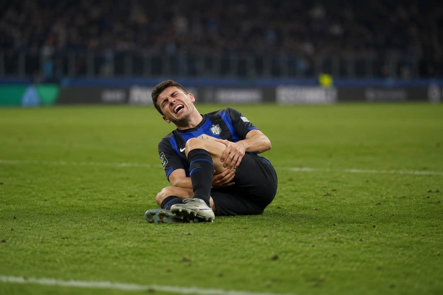
MUSCLE INJURIES
Pulled a hamstring sprinting? Took a knock to the thigh in a match? Both are muscle injuries, but they are not the same thing, and that difference shapes how long recovery takes and what you should be doing in the meantime.
Here is how we think about it, and what the evidence says.
The short version.
Most sport muscle injuries fall into one of three types, and the type, not just how much it hurts, is what tells us roughly how long recovery will take. Getting a precise diagnosis early saves weeks later.
Download the quick summary PDF of the full article from the link below.
THE FUNDAMENTALS
Two very different ways to injure a muscle
Before we talk about how bad an injury is, the first question we ask is simple: was there contact, or not? It sounds basic, but it sets the whole picture.
Direct injuries
A blow to the muscle: a tackle, a collision, a stray knee in five-a-side. The classic example is a dead leg.
Here is the catch. These often settle faster than people expect, and precisely because the pain eases quickly, they are the ones most likely to be rushed and re-aggravated.
Contact
Quicker healing
Often rushed
Indirect injuries
No contact at all. The muscle is overloaded from within, usually while sprinting, cutting, jumping, or decelerating hard.
This is what we see most often, and it is the more complex group, with its own grading that we walk through below.
Sprinting
Change of direction
Most common
WHERE IT HAPPENS
The muscles we see injured most
A handful of muscles take the brunt of it, mostly because of the load they absorb during sprinting, kicking, jumping, and fast changes of direction. See if one of these sounds familiar.
LOWER LIMB
Hamstrings
The most commonly injured muscle in sport. Classically, pulling up sharply near top speed, as the muscle works hard to slow the leg down at the end of each stride.
LOWER LIMB
Quadriceps (rectus femoris)
Common in kicking sports: the strain you feel striking a ball hard or accelerating into a sprint. A direct knock here is the classic dead leg.
LOWER LIMB
Medial gastrocnemius (calf)
Often called tennis leg. A sudden push off or lunge, for example reaching for a drop shot or driving off the back foot to sprint.
LOWER LIMB
Adductors (groin)
Common with quick changes of direction: stretching to reach a ball, or twisting and cutting off one leg. It has a habit of returning if it is not managed well.
UPPER LIMB
Biceps brachii (upper arm)
Less common, but seen in the gym and in climbing: typically a heavy or awkward lift where the arm is forced straight against a load.
Sound familiar?
If you can pin down the moment it happened, that mechanism is one of the most useful things you can bring to your first appointment.
GRADING THE INJURY
What actually decides how serious it is
Several grading systems exist, and clinicians do not always agree on which to use. But nearly all of them come down to the same two questions. 3
Where the damage sits?
In the muscle, at the fascia, at the muscle-tendon junction, or reaching into the tendon itself. The deeper into connective tissue it goes, the longer it takes to
heal. This is the single biggest factor in how we judge recovery time.
How much is involved?
Both the width (how much of the cross-section) and the length (how far it runs along the muscle) matter. A small, contained injury behaves very differently from an extensive one.
THE THREE TYPES
Type 1, 2, or 3: what each one means for you
Indirect injuries are usually grouped into three types, based on where along the muscle the damage sits. This is the part that surprises people most: the type matters more for recovery time than how painful day one felt.3
Type 1
Myofascial
At the junction of the muscle and its outer sheath, the fascia. Fascia heals quickly, so this is usually the fastest to recover from.
Type 2
Myotendinous
At the muscle-tendon junction, where load peaks during a contraction. The type we treat most often.
MOST COMMON
Type 3
Tendinous
Reaches into the tendon. Tendon has a poorer blood supply, so this is the slowest to heal and the one we treat most cautiously.
Different tissues heal at different speeds, and that is the main reason the three timelines differ. It is also worth saying that return to sport is not a single day but a continuum: a gradual, progressive process as strength and function are rebuilt. 2,3
How long recovery usually takes
Time after injury. Each bar runs from the start of rehabilitation through a graded return to sport, which happens progressively rather than on one fixed day.
Type 1 · Myofascial
Muscle meets fascia
Return to sport time
1/2 to 1 MONTH
Type 2 · Myotendinous
Muscle meets tendon
Return to sport time
1 to 2 MONTHS
Type 3 · Tendinous
Into the tendon
Return to sport time
2 to 4 MONTHS
The marker shows roughly when the injured tissue itself has healed, which is the main reason the timelines differ; full return comes later, as strength and function are rebuilt. These are typical guides, not guarantees: individual factors such as age, previous injuries, training level, general health, and normal individual variation all play a significant part. 2
REHABILITATION
The first weeks: what we actually do
Early on, the aim is not to rush. It is to give the tissue what it needs to heal well, in the right order. How fast we progress, and how much load and discomfort we allow, depends on which of the three types it is and where along the muscle it sits. 4
The load increases as we move left to right. The exact timing of each phase shifts with the type and location of the injury.
Phase 1
0 to 5 days
Protect, then start moving
Protect the area and settle the swelling, with compression and elevation. Ice can still help with pain in these early days: some debate exists about its effect on healing, but it remains a reasonable option for comfort. We are more cautious with anti-inflammatory medication and tend to hold off where we can, because that first wave of inflammation is actually part of how the muscle heals. Gentle active movement usually begins around day 2. 5
Phase 2
Week 1 to 3
Load, guided by pain and type
Load increases gradually, and how much discomfort we allow depends on the type: a little (up to about 4 out of 10) is acceptable for Type 1, we keep it minimal (around 2 out of 10) for Type 2, and we stay more cautious throughout for Type 3. Lengthening (eccentric) work can start earlier for Types 1 and 2, and is usually held back to around three weeks for Type 3. 4
Phase 3
Week 3+
Strength, power, and sport-specific work
The focus shifts to rebuilding real strength, power, and confidence: eccentric loading, plyometrics, and drills that look like your sport, progressed by what the tissue and the objective data are telling us, not by the calendar. 4
The marker shows roughly when the injured tissue itself has healed, which is the main reason the timelines differ; full return comes later, as strength and function are rebuilt. These are typical guides, not guarantees: individual factors such as age, previous injuries, training level, general health, and normal individual variation all play a significant part. 2
DIAGNOSIS AND MONITORING
Where scans fit in, and why we like ultrasound
Imaging helps us get the diagnosis right from day one. In the clinic, ultrasound does a lot of the heavy lifting. 1
Ultrasound is quick, done right there at your appointment, and lets us watch the muscle move in real time. It works best hand in hand with a thorough clinical examination: together they give the most accurate picture. That combination lets us pinpoint exactly where and how big the injury is, which guides how we load it, gives a realistic estimate of your return to sport, and lets us monitor healing as we go. Just as importantly, it flags straight away the cases that need onward referral, for example a larger tear, a collection of blood that needs attention, or a red flag that warrants further investigation. 1
MRI adds a more detailed cross-sectional view and is valuable for deeper or more complex injuries, so we reach for it when the picture calls for it. For most everyday decisions, though, a good clinical assessment combined with ultrasound tells us what we need to know. 1
RETURN TO SPORT
So, when are you actually ready?
There is no single magic test or date. The decision rests on objective markers rather than how the muscle happens to feel that day, and rushing it is one of the most common reasons injuries come back. 2
✓ Strength restored, not just matched to the other side but back to the level it should be, compared against normative data
✓ Full, pain-free range of movement
✓ Sport-specific tasks performed with control, confidence, and no pain
✓ Biological healing time respected, so we are not outpacing the tissue 2
What we measure
Objective strength and load data, side-to-side symmetry, comparison against normative benchmarks, and functional testing that mirrors your sport. Numbers we can track, repeat, and hold ourselves to. 1, 2
Where imaging sits
Useful for the diagnosis and for monitoring healing along the way. It is not usually the deciding factor for return to sport on its own; that always depends on the case. 1

All of this sounds familiar?
We are here to help. Book an assessment and start your recovery precisely and immediately, with diagnostic ultrasound and objective strength testing guiding every step.
Still unsure?
Book a free 15-minute discovery call. We will take a quick look, point you in the right direction, and help you work out the best next step.
Download the quick summary PDF of the full article from the link below.
References
- Guillodo, Y., Bouttier, R. and Saraux, A. (2011) ‘Value of sonography combined with clinical assessment to evaluate muscle injury severity in athletes’, Journal of Athletic Training, 46(5), pp. 500–504. Draghi, F., Zacchino, M., Canepari, M., Nucci, P. and Alessandrino, F. (2013) ‘Muscle injuries: ultrasound evaluation in the acute phase’, Journal of Ultrasound, 16(4), pp. 209–214.
- Pieters, D., Wezenbeek, E., Schuermans, J. and Witvrouw, E. (2021) ‘Return to play after a hamstring strain injury: it is time to consider natural healing’, Sports Medicine, 51(10), pp. 2067–2077.
- Macdonald, B., McAleer, S., Kelly, S., Chakraverty, R., Johnston, M. and Pollock, N. (2019) ‘Hamstring rehabilitation in elite track and field athletes: applying the British Athletics Muscle Injury. Classification in clinical practice’, British Journal of Sports Medicine, 53(23), pp. 1464–1473.
- Hickey, J.T., Timmins, R.G., Maniar, N., Rio, E., Hickey, P.F., Pitcher, C.A., Williams, M.D. and Opar, D.A. (2020) ‘Pain-free versus pain-threshold rehabilitation following acute hamstring strain injury: a randomized controlled trial’, Journal of Orthopaedic & Sports Physical Therapy, 50(2), pp. 91–103. Bayer, M.L., Magnusson, S.P. and Kjaer, M. (2017) ‘Early versus delayed rehabilitation after acute muscle injury’, New England Journal of Medicine, 377(13), pp. 1300–1301.
- Dubois, B. and Esculier, J.-F. (2020) ‘Soft-tissue injuries simply need PEACE and LOVE’, British Journal of Sports Medicine, 54(2), pp. 72–73. Morgan, C., Konopinski, M., Dunn, A. and Milsom, J. (2018) ‘Rehabilitation of rectus femoris injuries in kicking athletes’, Sport Performance & Science Reports, 1, 31.